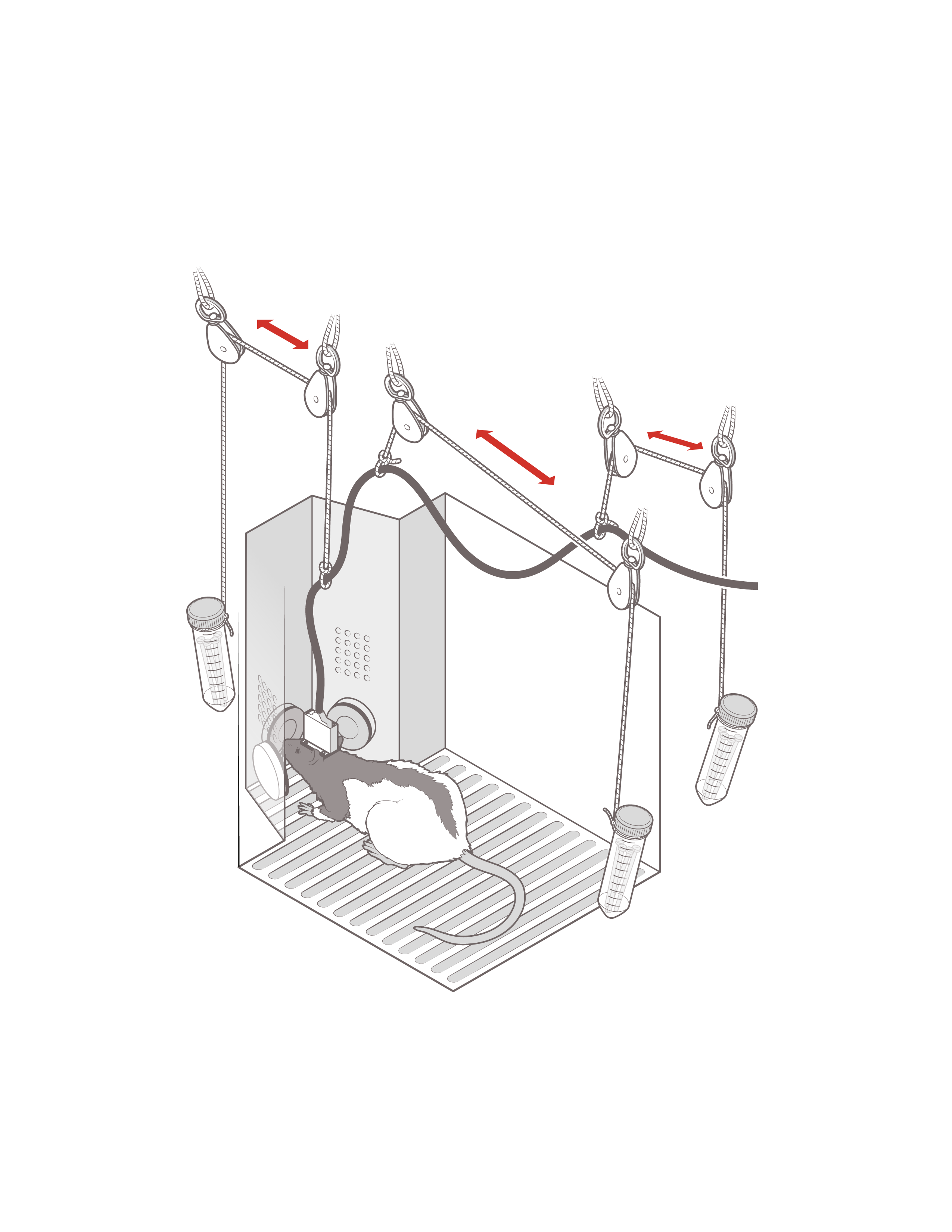
Surgical procedures for mounting the fUS system onto a rat.
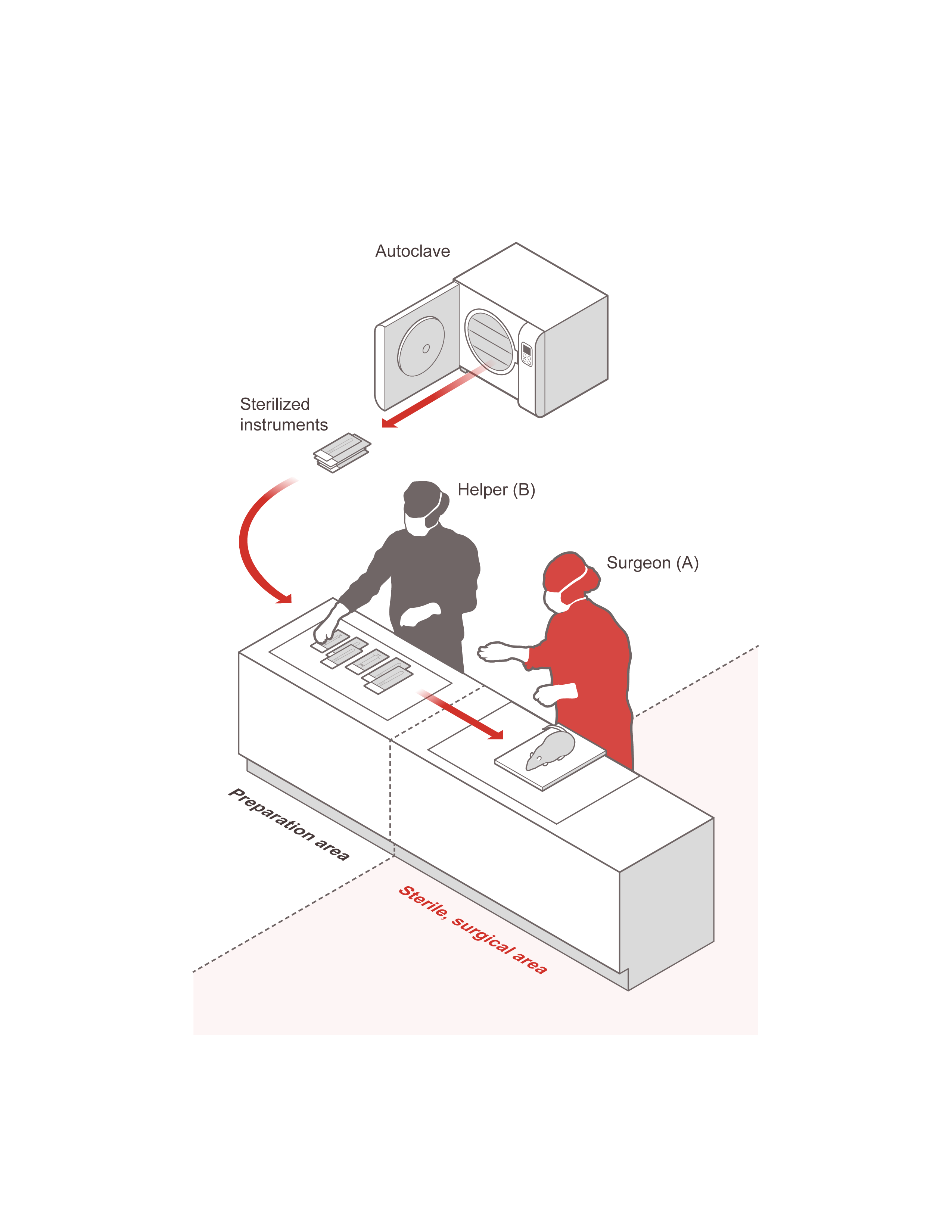
Surgery preparation. All tools used in the surgery should be sterilized and stored in sterile pouches. There are two areas: the preparation area where the helper B puts all the sterilized tools, the sterile surgical area where the surgeon A wearing sterile gloves take the sterile tools from the helper. This configuration ensures that the surgery is following a strict sterile procedure.
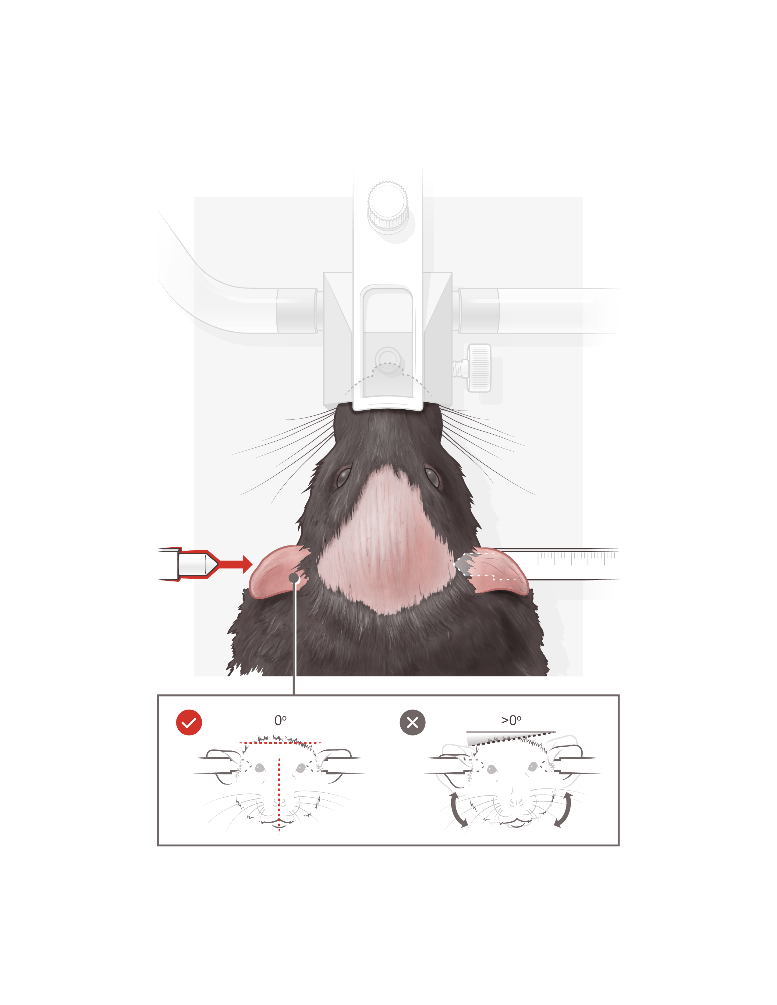
Ear bar insertion to stabilize the rat head. Note that the rat head is exposed after shaving ( the dark rose part in the middle). The shaving should be wide enough close to the ears and to the frontal side of the head. The inset shows the correct orientation of the head when inserted in the earbar.
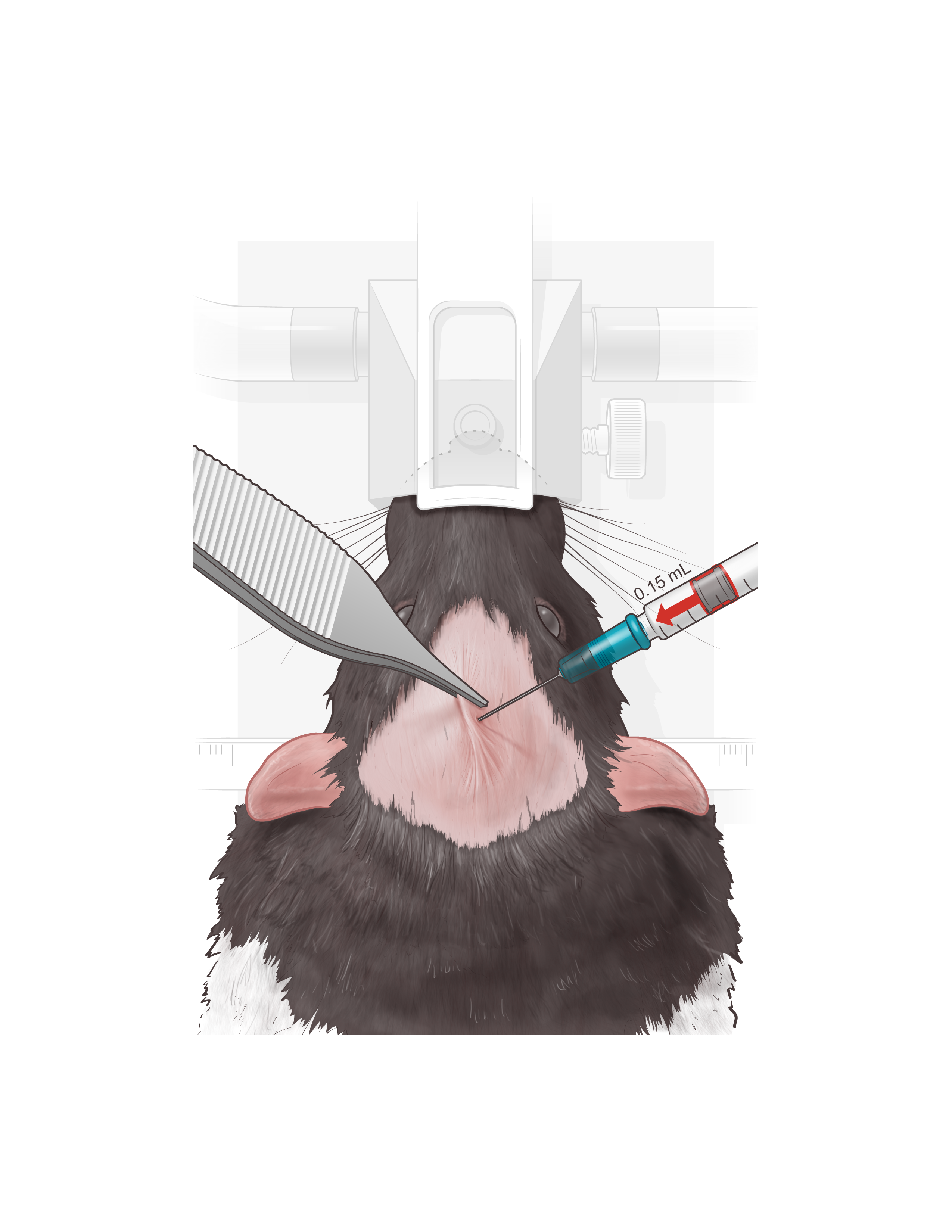
Lidocaine/Norepinephrine injection. Use a forceps to hold the skin to inject 0.15 ml lidocaine/norepinephrine mixture into the skin.
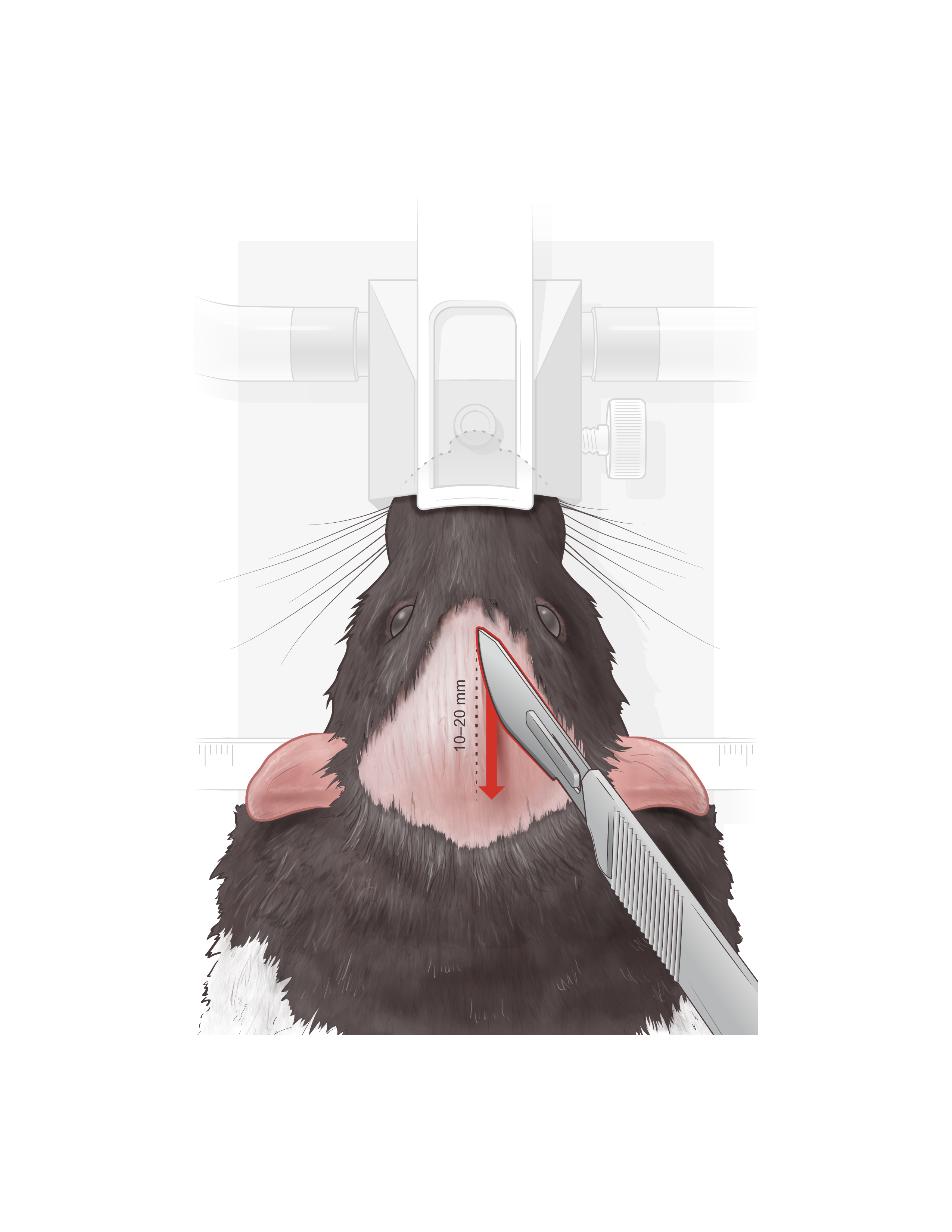
Use the scalpel to do a lateral incision in the skin varying between 10 - 20 mm depending on the intended size of the craniotomy to be performed afterwards
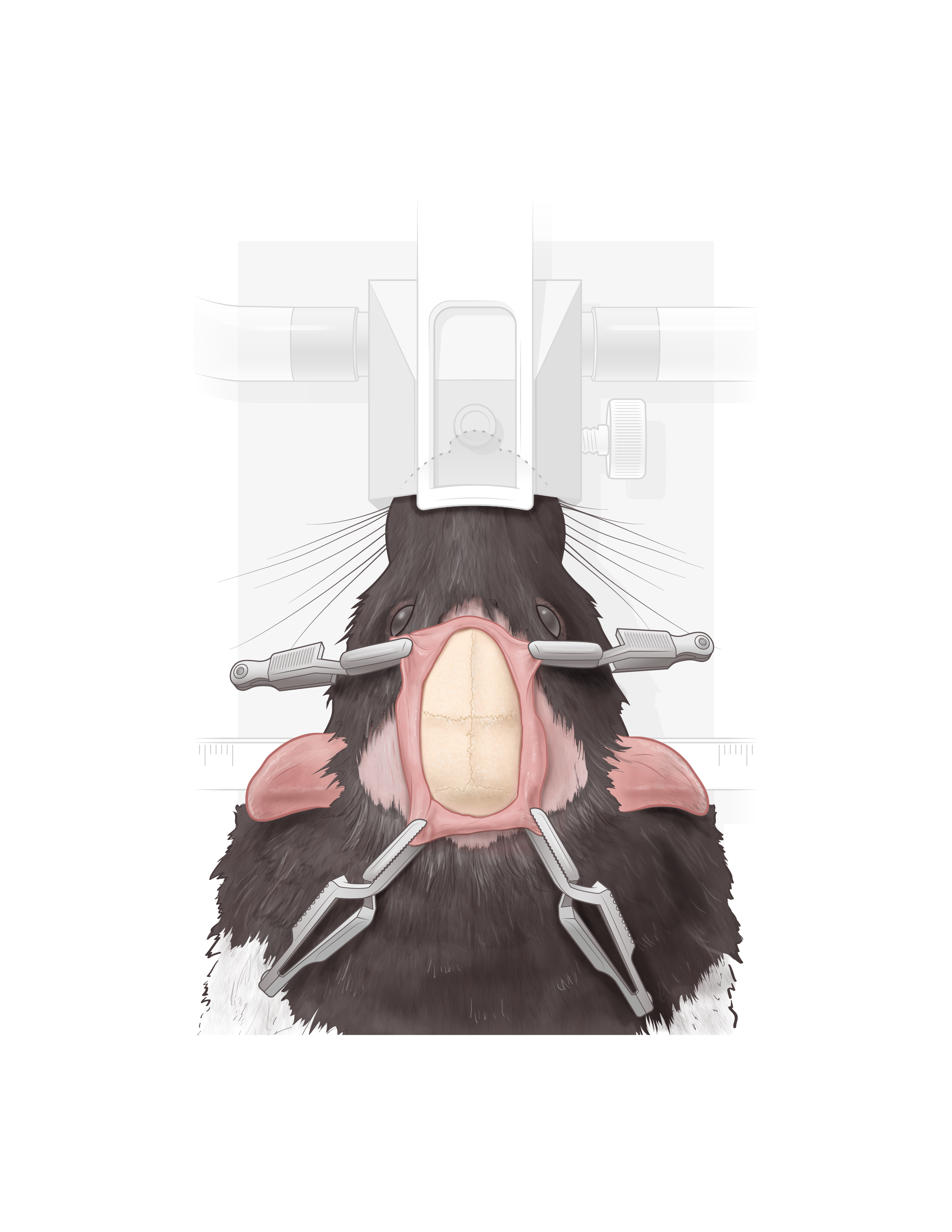
Using four surgical clips ,attached to the periosytum, to expose the skull and prevent the skin from folding over the skull. The skull appears clean after scrubbing away any remaining tissue debris or blood.
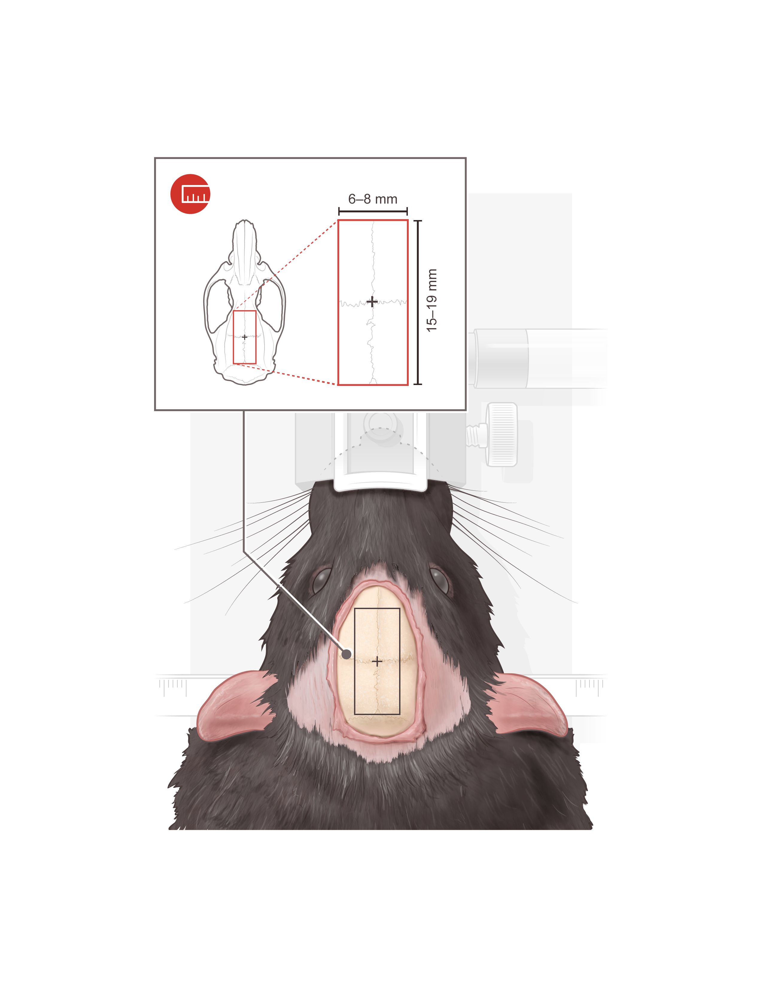
The surgical clips should remain there constantly but we remove them for clarity of presentation. The black rectangle represents the contours of the skull area in which the craniotomy will be performed. The black rectangle is marked and drawn using a sterile pen and a sterile waterproof ruler. The size of the rectangle can vary between 6 - 8 mm and 15 - 19 mm depending on the desired size of the craniotomy.
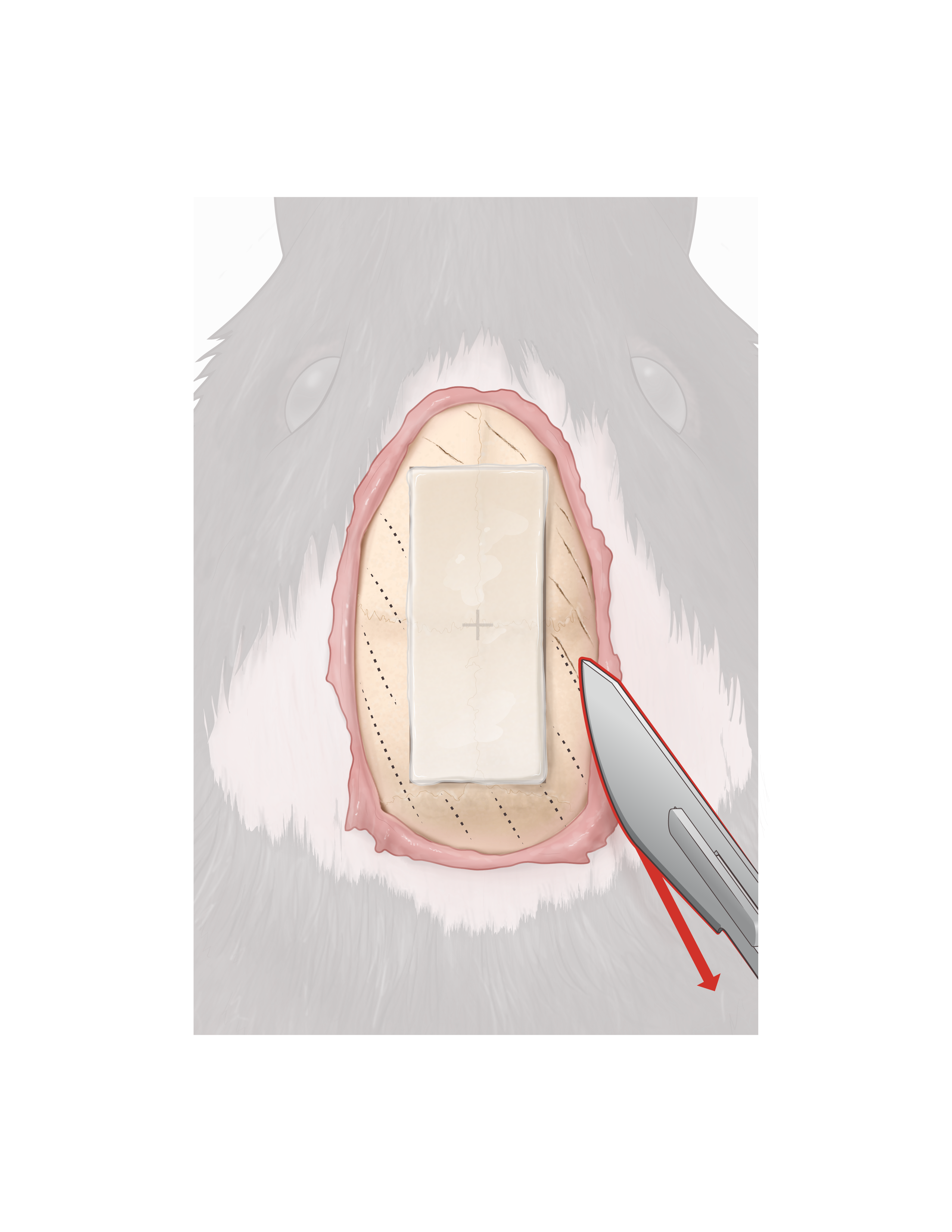
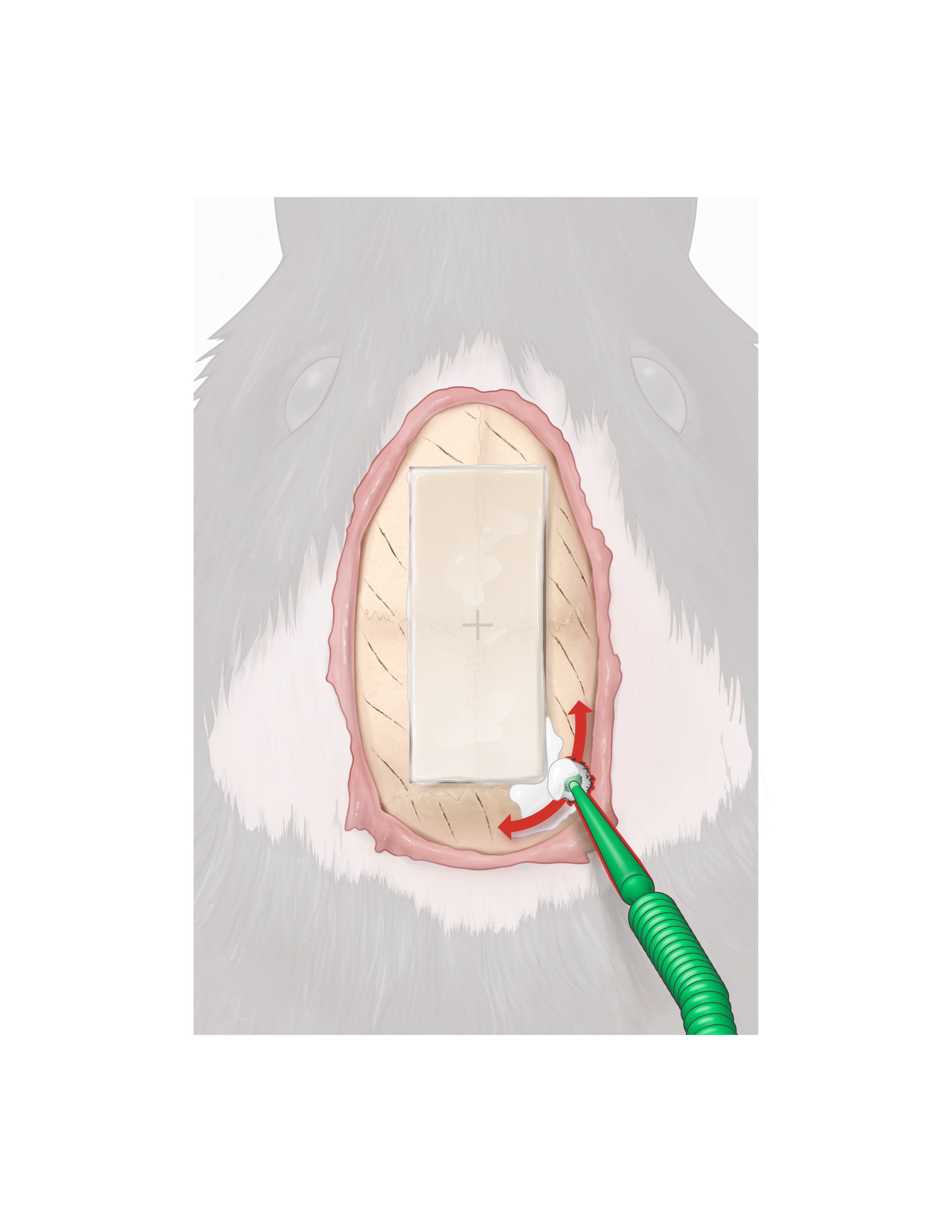
Kwik-sil is put on the rectangle carefully to fill in the black drawn rectangle then extras are cut using a scalpel to make sure that the rectangular gel piece is homogeneous and fits on the black rectangle marked on the skull. Using the scalpel one makes marks on the surface of the skull around the rectangular gel.
Using a sterile applicator, one puts copious amounts of metabond covering the skull surface around the rectangular gel piece.
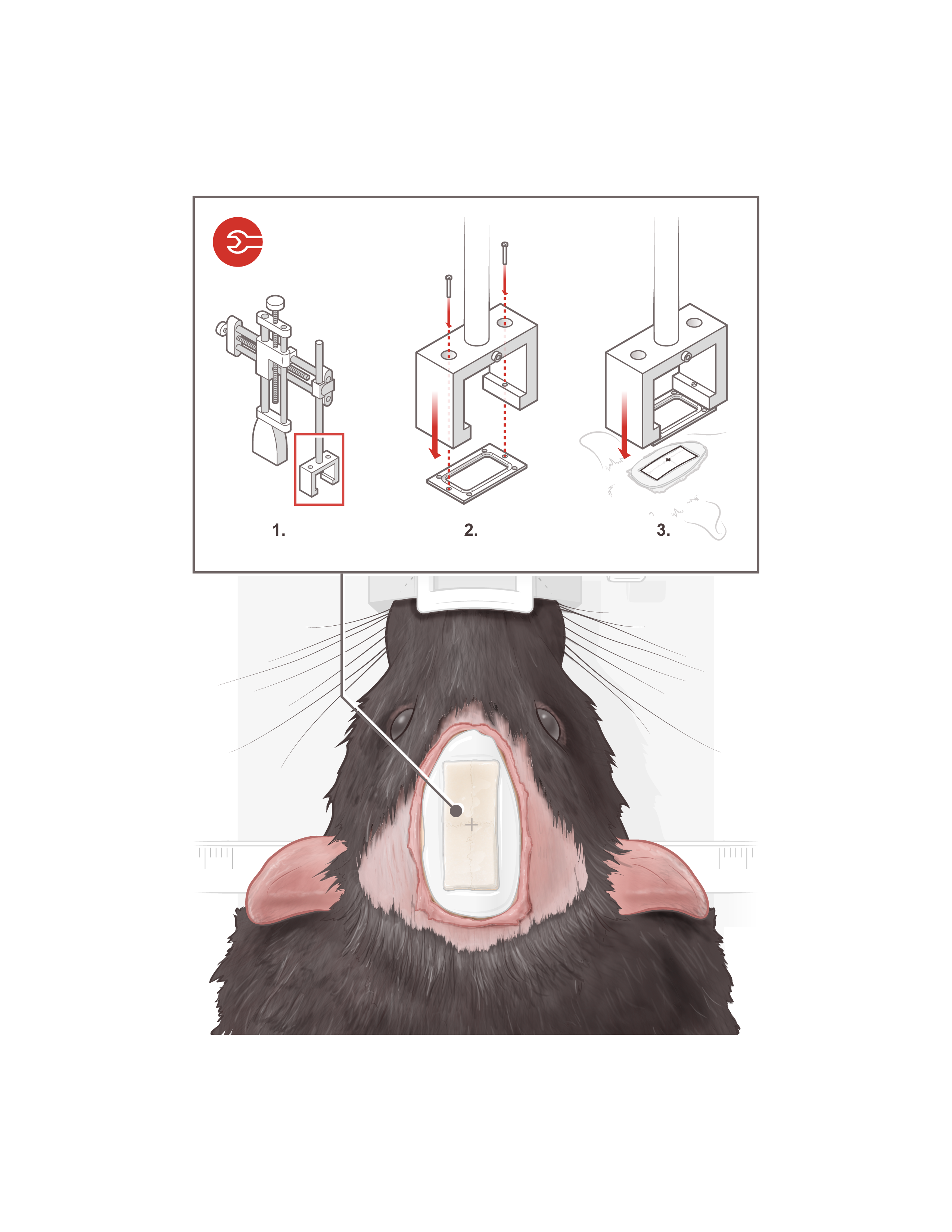
Inset: the headplate is screwed to the headplate positioning device attached to the stereotaxic positioning arm for precise manipulation. Subsequently it is lowered and centered carefully and precisely over the area where the rectangular gel is.
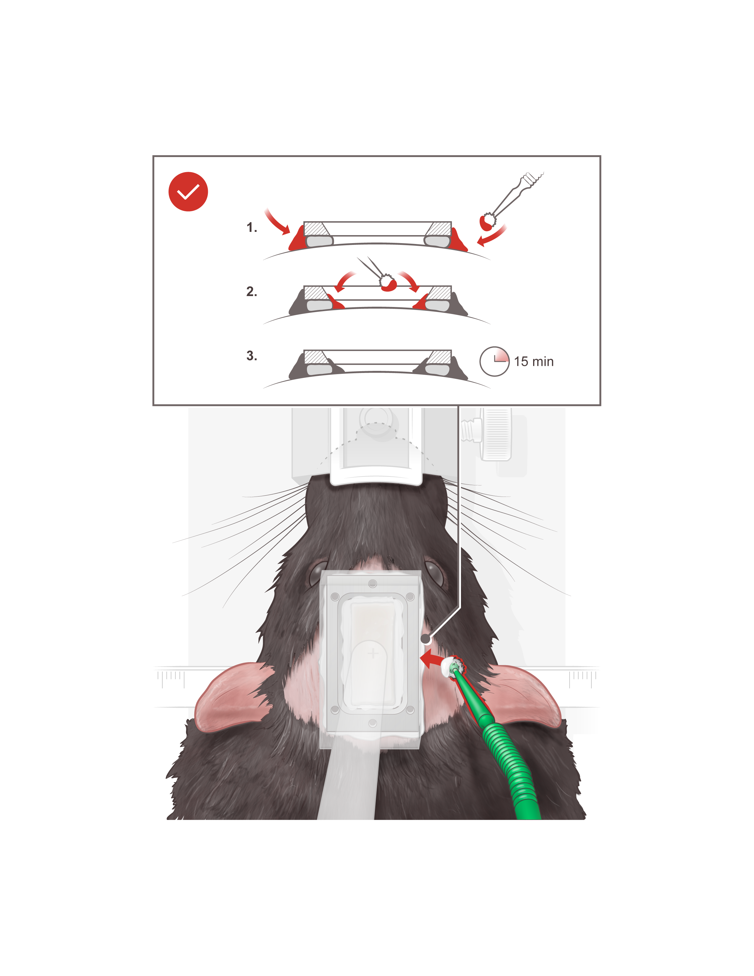
Using a sterile applicator metabond is applied. Metabond is applied on all sides and left for 15 minute. Then the rectangular gel over the skull is removed.
The headplate is then unscrewed from the headplate positioning system and lifted leaving the headplate firmly attached to the skull.
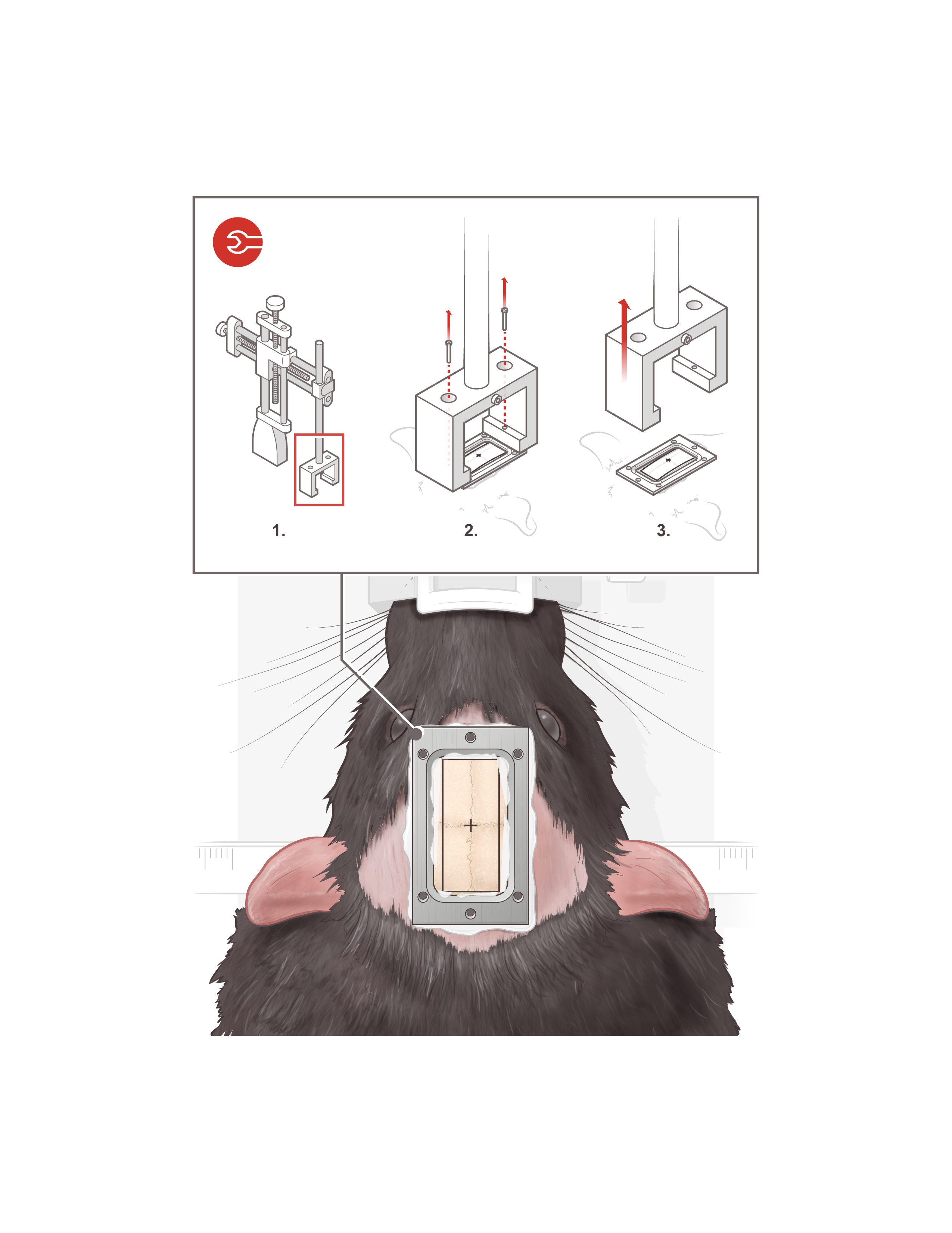
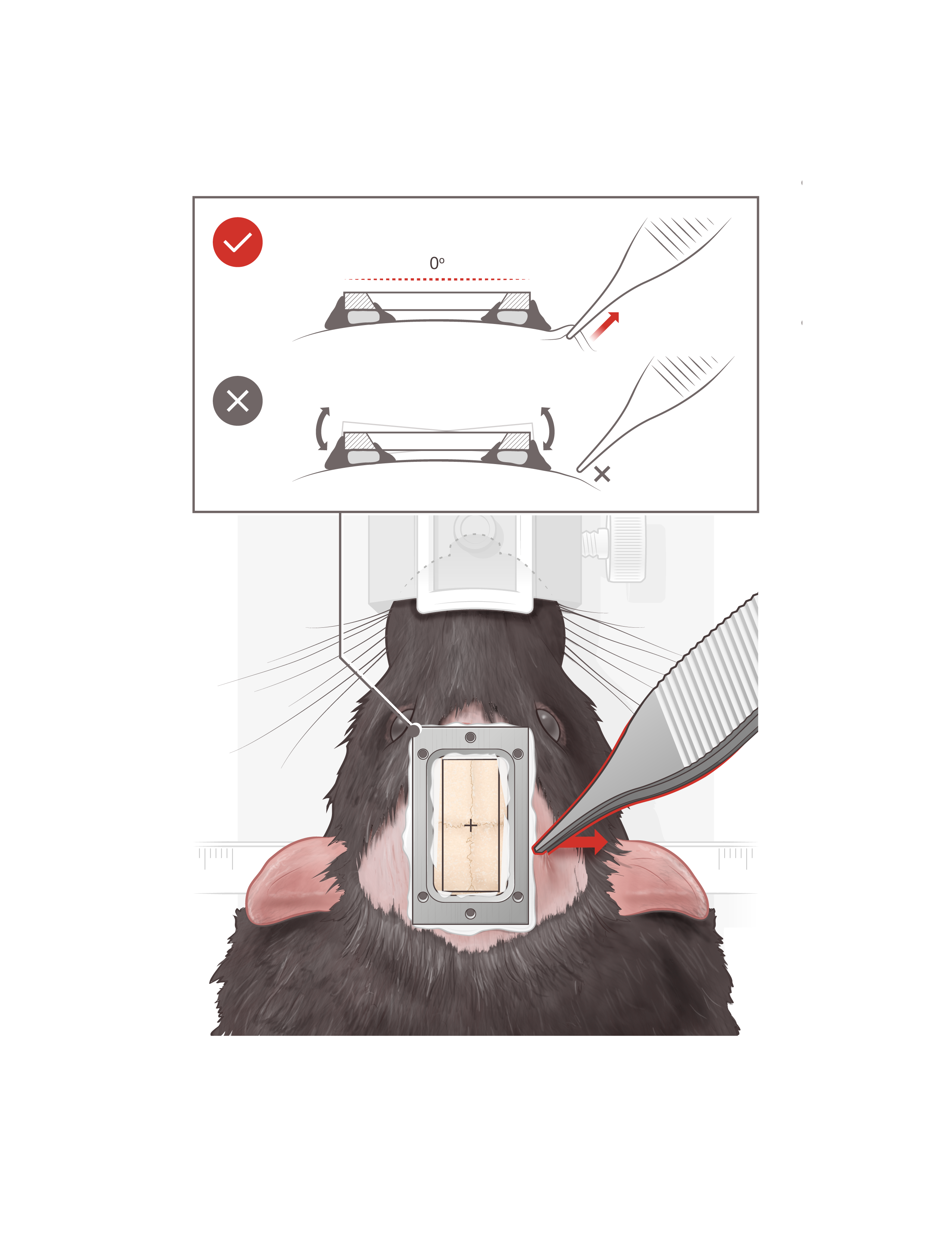
By pulling the skin using forceps, the headplate should be stable in a horizontal position and not move at all.
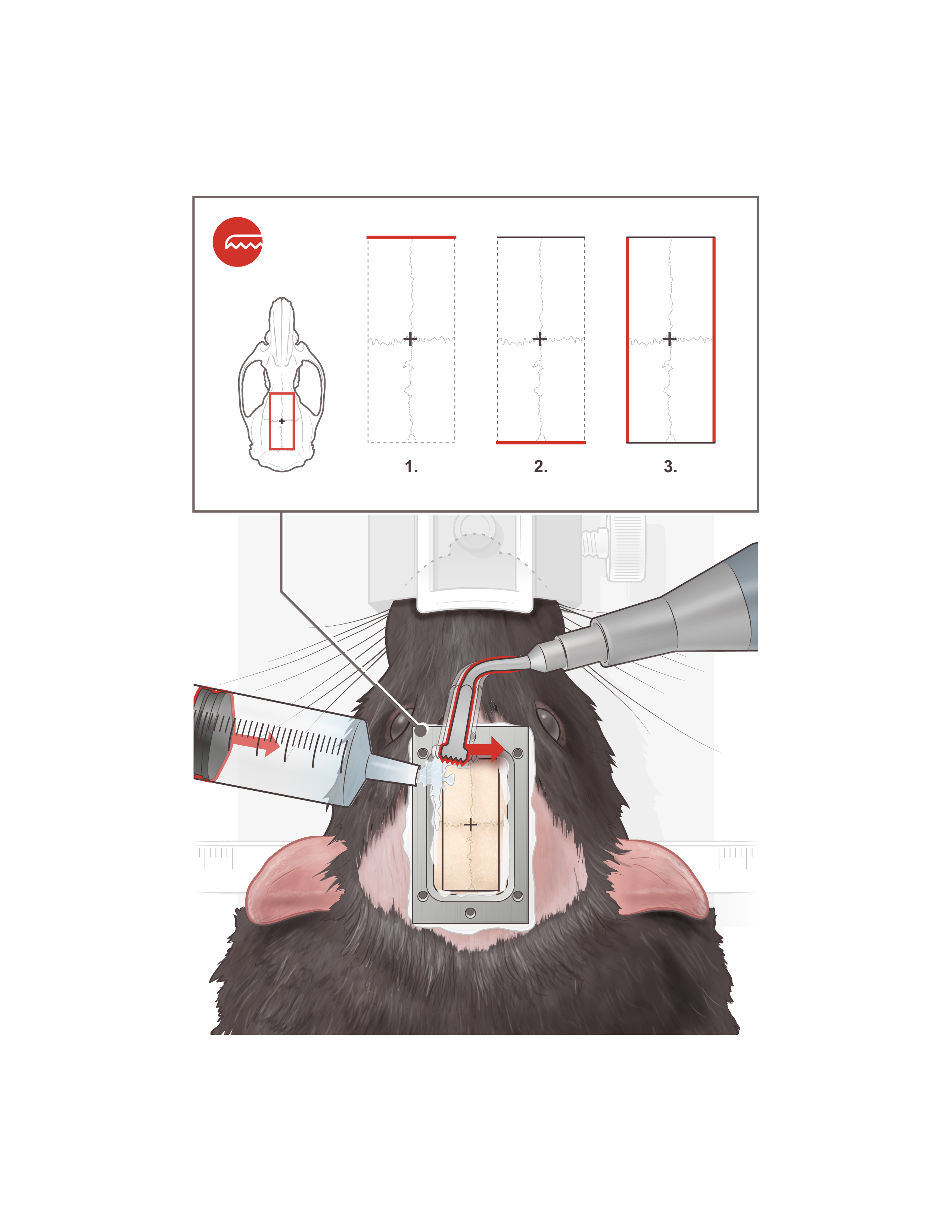
The craniotomy is performed using a piezo-electrical device called piezosurgery apparatus . The craniotomy is performed by beginning to cut , using a micro-saw insert, the upper part followed by lower part then the two sides of the cranial skull surface. To prevent overheating of the skull, sterile water is applied on the skull during the performance of the craniotomy.
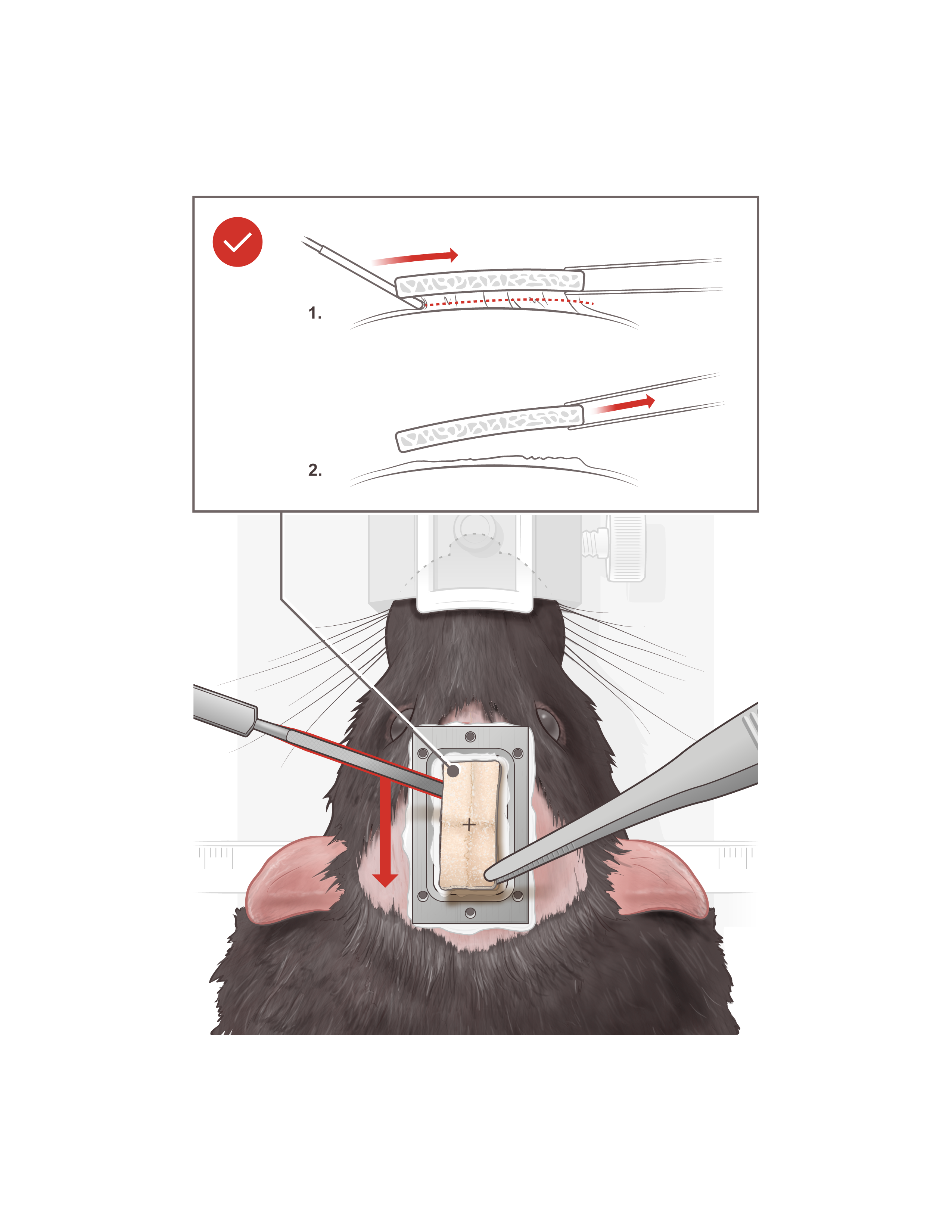
Using a forceps and a dissecting knife the cut skull after craniotomy is carefully removed very slowly.
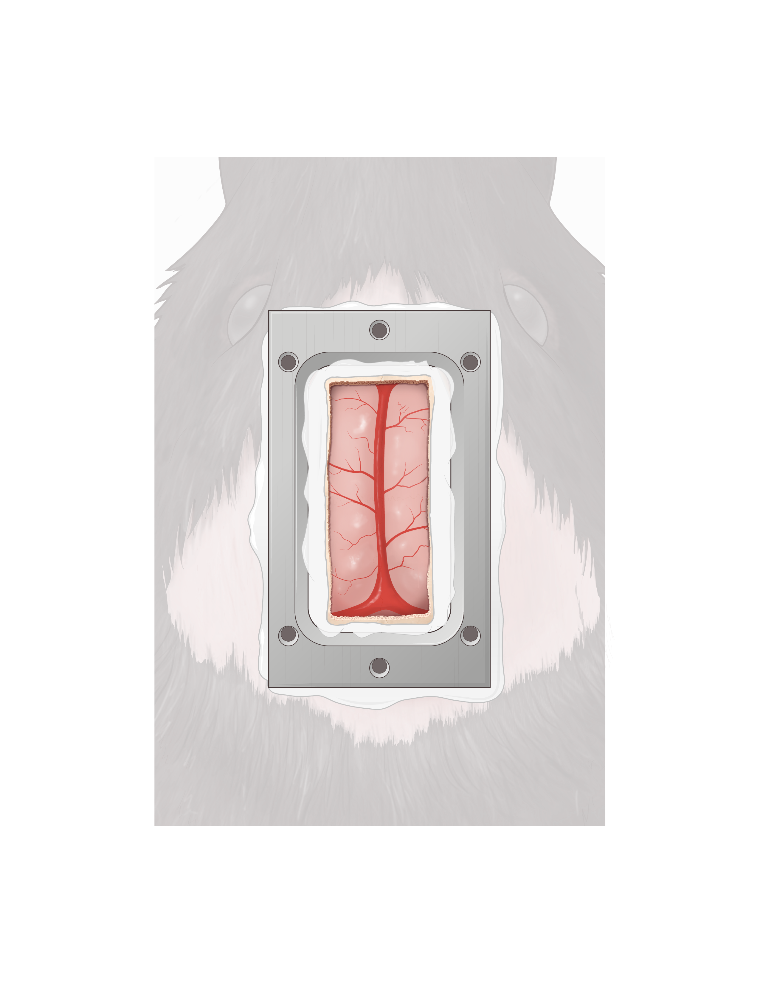
Exposed brain surface with the mid-saggital vein clear along the midline after the removal the skull piece.
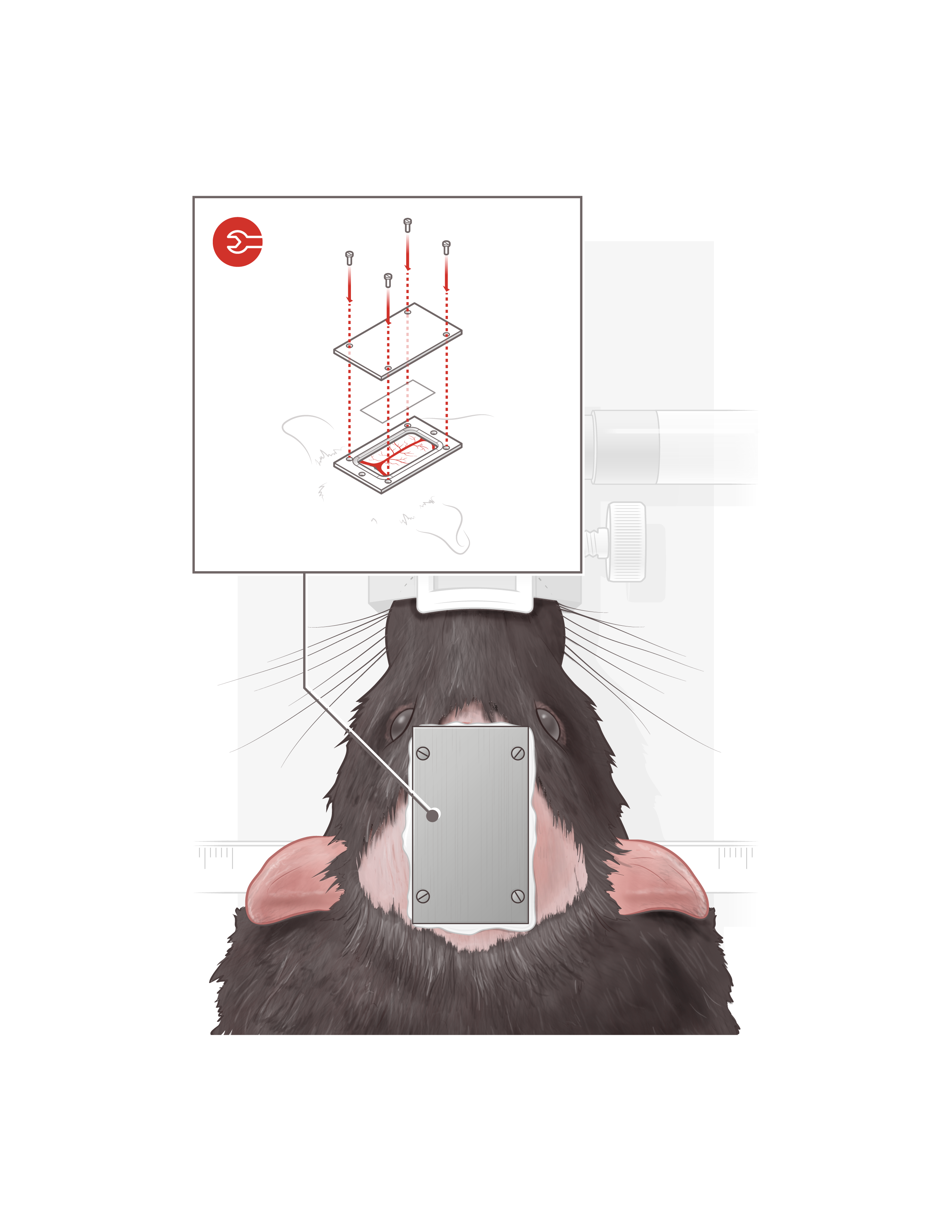
Polymer film (PMP) is put on the brain surface then headcover is screwed to the headplate.
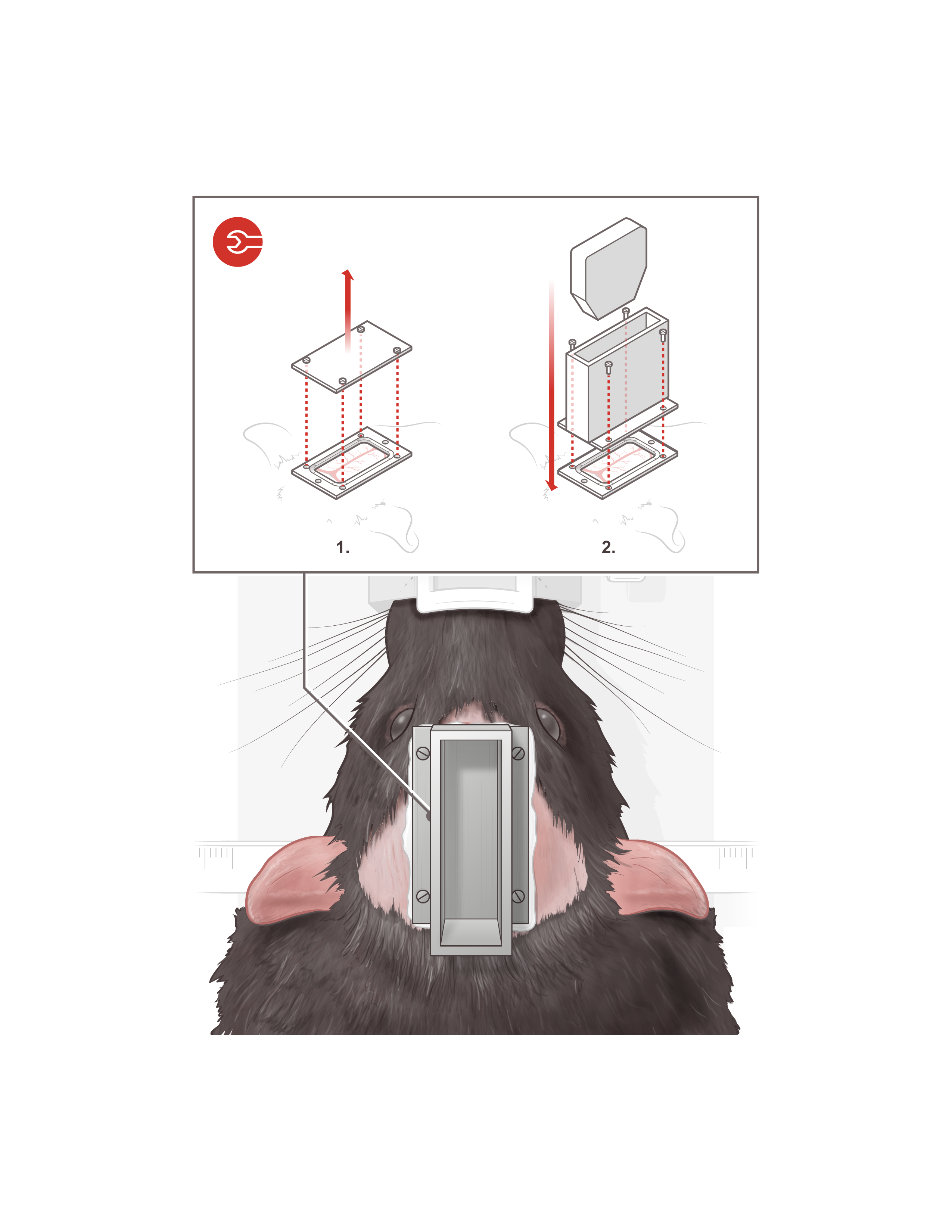
Headcover removed, probeholder attached to the headplate and the mock probe inserted.
Rat tethered with the functional ultrasound probe inside the behavior operant chamber.
